DM2
One repeat expansion. A slow unraveling of muscle function. Myotonic Dystrophy type 2 is a progressive disorder where toxic RNA silently disrupts the machinery that keeps muscle cells working.
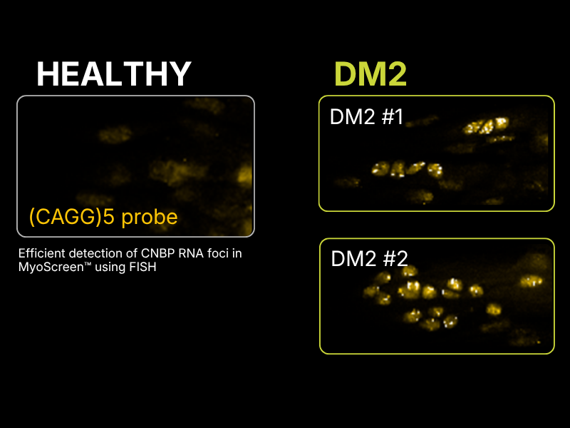
CYTOO’s MyoScreen™ platform models DM2 pathology in micropatterned primary human myotubes, enabling high-content detection of RNA foci, splicing dysregulation and downstream functional defects giving your program the cellular-level evidence it needs to move forward.
CYTOO's approach
At CYTOO, our MyoScreen™ platform leverages physiologically relevant micropatterned skeletal muscle cultures derived from human primary donor cells to model key cellular features of DM2.
This system enables high-content imaging combined with AI-based analysis to capture disease-relevant phenotypes, including altered myotube organization and maturation, nuclear RNA foci formation, mislocalization of splicing regulators, and defects in calcium handling and contractile performance.
These multiparametric readouts support the assessment of therapeutic strategies targeting toxic RNA, correction of splicing defects, and restoration of downstream muscle function.
Overall, CYTOO’s technologies provide a robust end-to-end platform to advance DM2 drug development.
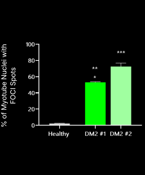
Data presented as mean ± SD Ordinary one-way ANOVA vs Healthy
About DM2
DM2 is a progressive, autosomal dominant genetic disorder affecting ~1 in 6,000–10,000 adults. It is caused by a CCTG repeat expansion in the CNBP (ZNF9) gene, leading to the accumulation of toxic RNA transcripts containing expanded intronic repeats. This RNA toxicity impairs RNA-binding proteins and disrupts alternative splicing regulation. Biological consequences include muscle weakness, myotonia, and abnormal metabolic signalling associated with reduced mitochondrial oxidative capacity and altered energy balance. Misplicing also affects transcripts encoding ion channels and muscle structural transcripts, contributing to defective Ca²⁺ handling and impaired contractility.
Explore our catalog of characterized donors and readouts
How can we work together
Everything is tailored to your needs through a flexible R&D partnership model that fosters true collaboration and innovation. We offer adaptable project structures and FTE allocation to fit your goals.