Master cell organization with CYTOO's micropatterns
Optimized solutions for research and high-throughput screening.
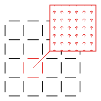
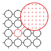
What is a micropattern?
Micropatterning is the miniaturization of adhesive patterns using lithography techniques. Widely used in cell biology, this technology allows precise control over cell adhesion, migration, and 2D/3D confinement. By restoring key mechanical properties such as geometry, architecture, composition, mechanics, and dynamics, micropatterning brings in vitro conditions closer to physiological reality.
CYTOO has developed extensive expertise in designing and producing optimized micropatterns for various cell types and research applications. Our advanced photolithography technology ensures exceptional precision and reproducibility.
Our solutions: two formats to fit your needs
Each cell type and application require a specific combination of shape, size, density, and coating protein for optimal results.
CYTOOchips
Designed for fundamental research and live-cell imaging, they can be used with dedicated accessories for an optimized experience.
CYTOOplates
Available in 96-well or 384-well formats, ideal for high-throughput screening.
FAQs
Find answers to the most frequently asked questions below. If you don’t find the information you’re looking for, please don’t hesitate to contact us!
Micropatterns
Does the geometry of the micropattern affect cell physiology?
Once you find the optimal size and pattern for your assays, the cells will not be impacted by the micropatterning. They will proceed through their normal cell division cycle at their usual pace.
To determine the best pattern for your needs, you can start with our Starter CYTOOchip. We also offer custom micropattern designs tailored to your specific requirements, such as patterns that promote cell-cell contacts.
What types of assays can be performed using micropatterns?
Simply replace your standard coverslip or glass-bottom microplate with a CYTOOchip and continue your usual cell assays with only minor adjustments. You can fix cells or record videos, and use your regular drugs, labels, and probes.
How do I select the ideal micropattern for my cell assay?
Start with our Starter CYTOOchip, which offers several standard micropattern geometries in several different sizes. Perform your cell assay on the Starter’s CYTOOchip and analyze which micropattern yields the best results. You can also refer to the description of micropattern shapes and sizes and their specific uses for additional guidance.
How can I tell if a micropattern is too small for my cells?
If the micropattern is too small, cells will not spread properly; they will maintain a rounded shape and appear bright in phase contrast. You might also observe the formation of blebs on the cells.
How can I tell if a micropattern is too large for my cells?
On larger micropatterns, cells will adopt various shapes, and in time-lapse microscopy, they will not stabilize on the pattern. Instead, they will exhibit significant movement back and forth. This leads to increased variability in cell phenotypes.
Precautions during use
My CYTOOchips are scratched: what could be wrong?
Although our products undergo strict quality control, if you notice scratches on your chips, please check the following:
- CYTOOchips should only be handled by the edges using tweezers. Avoid touching the central part of the CYTOOchip, as this can damage the cytophobic surface locally.
- When mounting the chips on glass, always use a mounting medium and ensure the chip stays wet during your experiments.
What should I do if my CYTOOchamber leaks?
Replace the gasket (spare gaskets are available for purchase from CYTOO).
Never use oil as it causes the gasket to swell and can lead to leaks.
Check that your CYTOOchambers have not been stored near strong magnetic fields, as this may demagnetize them.
Storage conditions
How should I store my CYTOOchips or CYTOO plates?
Store our products refrigerated at 4°C in their original packaging.
Do not freeze the plates, as this could cause the glass bottom to detach from the upper frame.
How long can I store my CYTOOchips or CYTOOplates?
Use the products before the expiration date indicated on the blister and bag. Once the bag is opened, use the chips and plates promptly, as optimal storage conditions are no longer guaranteed.
How should I store my CYTOOchambers?
Store our products refrigerated at 4°C under dry conditions in its original packaging. Do not freeze the plates, as this could cause the glass bottom to detach from the upper frame. Avoid storing CYTOOchambers near strong magnetic fields (e.g., solenoids or high-content screening systems), as this may demagnetize them and cause leaks. CYTOOchambers can be autoclaved, but do not heat them above 120°C as a precaution.