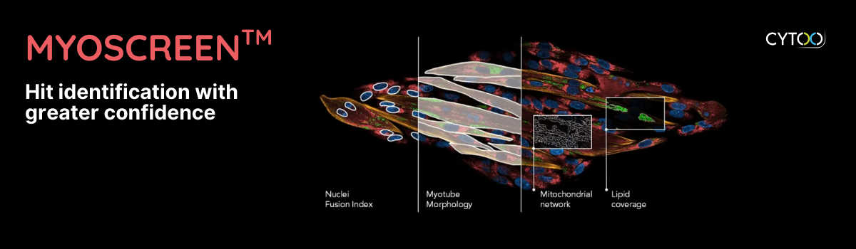
What we capture in MyoScreen™ healthy AND diseased myotubes:
✨ Morphology
✨ Integrity & mass
✨ Membrane potential
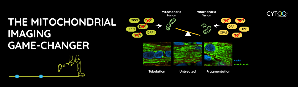
Our triple-approach strategy:
🎯 Texture analysis (10X magnification) → High-throughput screening of patterns (spots, edges, ridges, dark regions)
🔬 Branch length (40X magnification) → Precise fusion/fission balance measurement
🤖 AI-powered Mitoprofiling (40X magnification) → Catch distinct mitochondrial phenotypes, including subtle or mixed populations
We are expanding our readouts: What additional mitochondrial readouts or functions would you most like to evaluate in myotubes through fluorescence imaging?