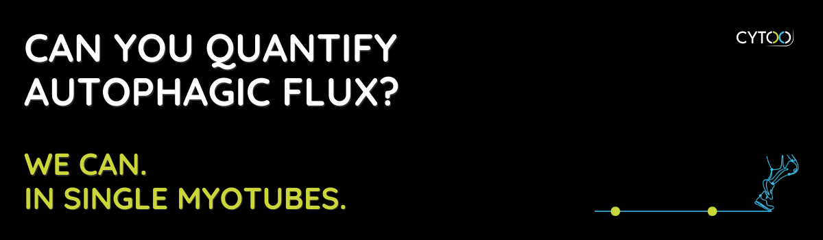
Our MyoScreen platform addresses this gap.
Through advanced fluorescence microscopy and high-content analysis of individual myotubes, we enable:
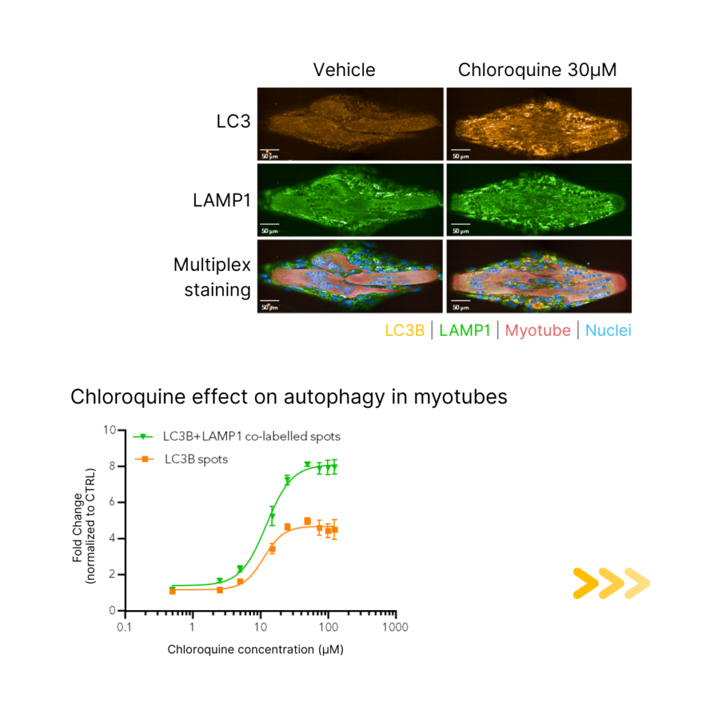
✔️ Sensitive visualization of LC3B-positive autophagosomes
✔️ Precise identification of LC3B/LAMP1 co-labelled autolysosomes
✔️ Robust quantification of autophagic flux
✔️ Simultaneous assessment of myotube size, differentiation & viability
This integrated approach supports high-throughput screening for compounds that modulate autophagy — whether the goal is to enhance it (e.g., myofibrillar myopathies) or inhibit it (e.g., Pompe disease).
📊 See image: Chloroquine treatment reveals dose-dependent accumulation of autophagic compartments, readily quantified by MyoScreen.