
Muscle wasting Disease
Four signaling axes. One shared endpoint. The progressive loss of muscle that drives cachexia, sarcopenia, and metabolic disease.
CYTOO’s MyoScreen™ platform enables quantitative assessment of skeletal muscle atrophy and rescue in human primary myotubes, supporting sensitive, high-throughput evaluation of muscle-preserving and regenerative compounds.
CYTOO's approach
At CYTOO, our MyoScreen™ platform relies on physiologically
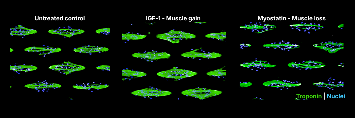
Leveraging this model, high-content imaging and analysis deliver highly sensitive readouts — myotube area, fusion index, and nuclei count — that capture the full range of skeletal muscle responses, from atrophy to preservation and regeneration.
Leveraging this model, high-content imaging and AI-based analysis deliver highly sensitive readouts — myotube area, fusion index, and nuclei count — that capture the full continuum from muscle loss and atrophy to preservation and regeneration.
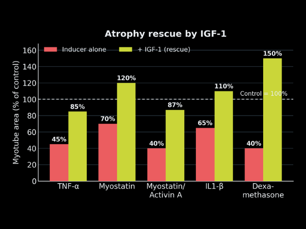
Crucially, our atrophy models are rescue-ready: candidate compounds are evaluated by the percentage recovery of myotube mean area and fusion index — a stringent, translatable readout. We offer flexible protocol designs, applying treatments either during myoblast differentiation or on fully differentiated myotubes, to best capture a compound’s mode of action and treatment window.
Overall, CYTOO’s technologies offer an end-to-end platform spanning the muscle-wasting drug development pipeline, from early candidate identification to advanced potency assay development supporting preclinical and clinical programs.
About Muscle Wasting Disease
Skeletal muscle homeostasis relies on a balance between protein synthesis, degradation, and regenerative capacity. Disruption of this balance underlies conditions such as cancer cachexia, sarcopenia, type 2 diabetes, and over-accelerated weight loss.
Muscle wasting is driven by four main signaling axes: suppression of the IGF-1/Akt/mTOR pathway; activation of TGF-β family signaling (myostatin…) via Smad2/3; increased pro-inflammatory cytokine signaling (TNF-α…) through NF-κB and STAT3; and enhanced glucocorticoid signaling inducing atrogene expression (MuRF1, Atrogin-1) — collectively driving an upregulation of the ubiquitin-proteasome system and autophagy.
Our assay suite evaluates the full spectrum, from muscle loss and atrophy rescue to muscle preservation and regeneration, using primary human skeletal muscle cells from healthy and/or diseased donors spanning a range of ages and biological sexes — validated for responsiveness to the four key signaling pathways regulating muscle differentiation and loss on our MyoScreen™ platform.
Explore our catalog of readouts
How can we work together
Everything is tailored to your needs through a flexible R&D partnership model that fosters true collaboration and innovation. We offer adaptable project structures and FTE allocation to fit your goals.